A modern eye clinic depends on more than a row of impressive instruments. It needs reliable tools that help clinicians assess vision, examine eye structures, and track changes over time. From the cool chin rest of an autorefractor to the focused beam of a slit lamp, each device plays a practical role. The right Optometry Equipment supports consistent measurements, but it does not replace clinical judgment, patient history, or clear communication. Small details matter.
This guide introduces ten common equipment types found in eye clinics, including visual acuity charts, phoropters, autorefractors, slit lamps, tonometers, lensometers, retinal cameras, optical coherence tomography systems, visual field analyzers, and trial lens sets. Each tool answers different clinical questions. A tonometer measures eye pressure, while a retinal camera captures images that can support documentation and follow-up. Some equipment works together during an examination; other tools are used only when a patient’s needs call for them.
Choosing equipment is not simply a matter of buying the newest model. Clinics may need to weigh patient volume, available space, staff training, maintenance, and compatibility with existing systems. Calibration and careful operation matter, too. A crowded room can slow appointments, while an under-equipped room may limit available assessments. There is no universal setup. This overview offers a useful starting point, though local workflows and clinical needs should guide the final choices.
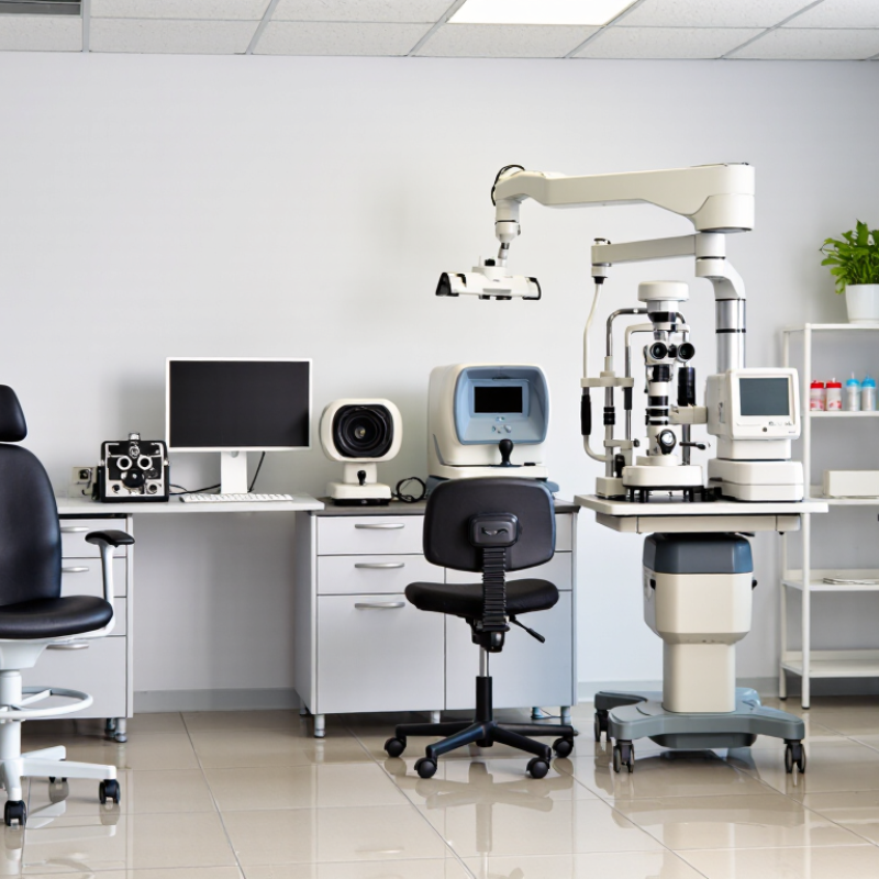
Visual acuity and refraction testing equipment helps clinicians measure how clearly a patient sees and identify possible focusing errors. A wall chart or calibrated digital display presents letters or symbols at a measured distance, often six metres or an optical equivalent. Consistent lighting matters; glare or dim rooms can affect responses. Keep the chart unobstructed and confirm the patient covers one eye without pressing on it. Small details matter.
For refraction, a phoropter or trial-frame lens set lets the clinician compare lenses while the patient reads the chart. A retinoscope provides an objective estimate by observing light reflected from the eye. An autorefractor can offer another estimate, but its reading is not a complete prescription. Patient feedback and clinical judgment remain essential. A child, for example, may need picture symbols or extra time to answer reliably.
Equipment needs regular checks for alignment, cleanliness, and calibration according to its instructions. Even a well-maintained room can produce inconsistent results if the testing distance changes. One limitation is easy to overlook: chart scores depend partly on attention, language, and fatigue. A number on the screen cannot explain every difficulty a patient reports.
A slit-lamp microscope gives clinicians a magnified, illuminated view of the eye’s front structures. The patient rests their chin and forehead against the supports while the examiner adjusts the beam and focus. Small movements matter. A narrow beam can reveal corneal layers and the depth of the anterior chamber; a wider beam helps inspect the iris and lens. With fluorescein dye and a cobalt-blue filter, surface staining may become easier to see. Findings still need clinical interpretation, not just a clear image.
Comfort affects the examination. Check that the chin rest is clean, the supports are stable, and the light is not unnecessarily bright. Explain where the patient should look before changing the beam. It sounds minor, but unclear instructions can make the view harder to maintain. One easy detail to overlook is alignment; a slightly off-center beam may hide the feature being examined.
An ophthalmoscope helps assess the fundus, including the optic disc, retinal vessels, and background retina. Direct instruments offer a close, upright view through the pupil, while indirect systems provide a broader view with different depth perception. Room lighting, pupil size, and the examiner’s technique all affect what can be seen. A limited view is not a normal result. Record the conditions and consider whether another examination method or referral is appropriate. Regularly check the optics and illumination; a dim or poorly focused instrument can obscure useful detail.
Tonometers help clinicians estimate intraocular pressure (IOP), an important part of assessing eye health and glaucoma risk. In a busy clinic, the choice often depends on measurement method, patient comfort, and workflow. Applanation tonometry measures the force needed to flatten a small area of the cornea. Rebound devices use a brief probe contact, while non-contact instruments measure pressure using a controlled air pulse. Each method has practical trade-offs. Small details matter. A patient squeezing their eyelids, holding their breath, or sitting awkwardly may affect a reading. Clean contact surfaces and routine equipment checks support reliable measurements.
IOP is one part of an eye examination, not a diagnosis by itself. Corneal thickness, time of day, medication use, and earlier readings can provide useful context. When a result seems unexpected, clinicians may repeat the measurement and check the patient’s position and technique. Repeat if needed. Staff training matters as much as equipment selection; in practice, this step can get rushed, and that deserves attention. A chart entry should record the method used, since comparing results from different methods may be misleading. No device is perfect, and a single reading rarely tells the whole story.
Retinal imaging gives clinicians a view of the back of the eye without relying only on a patient’s description. A clear image can document the optic nerve, blood vessels, and central retina for later comparison. Good positioning matters: a small head movement or an unfocused capture may hide useful detail. Image quality is not a diagnosis. Clinicians interpret findings alongside symptoms, examination results, and relevant health history.
Corneal mapping measures the shape and curvature of the cornea. Its color-coded maps can help assess irregular astigmatism and support contact lens fitting or pre-procedure evaluation. A dry eye can distort the surface and produce a misleading pattern. Repeat the scan when results do not fit the examination. Small details matter. Even then, maps need clinical interpretation; they should not be treated as a standalone answer.
Visual field devices measure how well a person detects small points of light in different parts of their vision. The patient watches a central target and responds when a light appears, often with a handheld button. Instructions and a quiet room help, but fatigue or uncertainty can affect responses. A few missed lights may reflect attention, not change in vision. Reviewing reliability indicators and comparing repeat tests helps clinicians judge whether a result is meaningful. In a busy clinic, this step can be rushed. It deserves care.
| Equipment | Primary Function | Typical Information or Output | Common Clinical Role |
|---|---|---|---|
| Slit-Lamp Biomicroscope | Provides magnified, illuminated views of the anterior eye. | Visual examination of the eyelids, cornea, conjunctiva, anterior chamber, and lens. | Routine assessment of eye structures and evaluation of signs such as inflammation, corneal changes, or cataract. |
| Autorefractor and Keratometer | Estimates refractive error and measures the curvature of the cornea. | Objective refraction estimates and corneal curvature readings, commonly used as starting measurements. | Supports refraction and contact lens assessments; results are generally refined with subjective testing. |
| Phoropter | Allows lenses to be changed during subjective refraction. | Patient responses used to determine a spectacle prescription, including sphere, cylinder, and axis. | Helps clinicians refine vision correction based on which lens options provide clearer vision. |
| Visual Acuity Chart | Measures the clarity of central vision at a specified testing distance. | Visual acuity recorded using standardized optotypes and notation, such as Snellen or logMAR. | Used in routine eye examinations and to document vision with or without correction. |
| Tonometer | Measures intraocular pressure (IOP). | Pressure readings, typically reported in millimeters of mercury (mmHg). | Provides one part of glaucoma assessment; pressure readings are interpreted alongside other clinical findings. |
| Fundus Camera | Captures photographs of the retina and optic nerve at the back of the eye. | Color or specialized retinal images for documentation and comparison over time. | Supports retinal screening and monitoring of visible retinal or optic nerve changes. |
| Optical Coherence Tomography (OCT) Scanner | Produces cross-sectional images of retinal and, on suitable systems, optic nerve structures. | Layer-based scans and measurements, including retinal thickness maps and optic nerve analyses. | Helps assess and monitor structural changes associated with retinal and optic nerve conditions. |
| Corneal Topographer or Tomographer | Maps corneal shape; some systems also assess the cornea's anterior and posterior surfaces. | Color-coded curvature or elevation maps and related corneal measurements. | Used in contact lens fitting, assessment of corneal irregularity, and preoperative evaluation. |
| Automated Visual Field Analyzer (Perimeter) | Measures sensitivity across a patient's visual field. | Threshold sensitivity results and visual field plots showing areas of reduced response. | Supports evaluation and monitoring of visual field loss, including changes associated with glaucoma. |
| Lensmeter (Lens Analyzer) | Measures the optical power of spectacle lenses. | Lens power, cylinder axis, and prism measurements when present. | Checks existing spectacles and verifies finished lenses against a prescribed correction. |
A lens analyzer earns its place by checking what a prescription lens actually delivers. It measures sphere, cylinder, axis, and often prism, helping staff spot a swapped lens or an incorrect axis before dispensing. Small details matter. A scratched lens or a poorly centered reading can distort results, so clean the lens and align its optical center carefully. Then verify unusual readings by repeating the measurement and comparing them with the prescription.
The World Health Organization’s 2019 World Report on Vision estimates that at least 2.2 billion people have near or distance vision impairment; at least 1 billion cases could have been prevented or remain unaddressed. The CDC reports that more than 12 million U.S. adults aged 40 and older have vision impairment, including about 8 million with uncorrected refractive error. These figures underline why reliable refraction tools matter. An autorefractor provides a starting estimate, while a phoropter helps refine subjective responses. A slit lamp supports examination of the eye’s front structures, and a tonometer measures intraocular pressure. It is easy to over-trust a tidy digital reading. Instruments need routine checks, and clinicians still interpret results in context. A rushed workflow can miss that.